
Researchers from Switzerland and Japan have now investigated this virus in minute detail. Using a microscopy technique that they developed themselves, the scientists can zoom in on the surface of human cells in a Petri dish. For the first time, this has allowed them to observe live and in high resolution how influenza viruses enter a living cell.
Led by Yohei Yamauchi, Professor of Molecular Medicine at ETH Zurich, the researchers were surprised by one thing in particular: the cells are not passive, simply allowing themselves to be invaded by the influenza virus. Rather, they actively attempt to capture it. “The infection of our body cells is like a dance between virus and cell,” says Yamauchi.
Viruses surfing on the cell surface #
Of course, our cells gain no advantage from a viral infection or from actively participating in the process, the researchers said. The dynamic interplay takes place because the viruses commandeer an everyday cellular uptake mechanism that is essential for the cells. Specifically, this mechanism serves to channel vital substances, such as hormones, cholesterol or iron, into the cells.
Once the cell’s receptors detect that a virus has attached itself to the membrane, a depression or pocket forms at the location in question. This depression is shaped and stabilised by a special structural protein known as clathrin. As the pocket grows, it encloses the virus, leading to the formation of a vesicle. The cell transports this vesicle into its interior, where the vesicle coating dissolves and releases the virus.
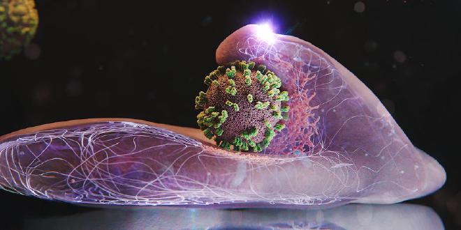
For the first time, researchers have observed live and in high resolution how influenza viruses infect living cells. This was possible thanks to a new microscopy technique, which could now help to develop antiviral therapies in a more targeted manner. Credits: Nicole Davidson / ETH Zurich
Previous studies investigating this key process used other microscopy techniques, including electron microscopy. As these techniques entailed the destruction of the cells, they could only ever provide a snapshot. Another technique that is used – known as fluorescence microscopy – only allows low spatial resolution.
Combined techniques, including for other viruses #
The new technique, which combines atomic force microscopy (AFM) and fluorescence microscopy, is known as virus-view dual confocal and AFM (ViViD-AFM). Thanks to this method, it is now possible to follow the detailed dynamics of the virus’s entry into the cell.
The new technique therefore provides key insights when it comes to the development of antiviral drugs. For example, it is suitable for testing the efficacy of potential drugs in a cell culture in real time. The study authors emphasise that the technique could also be used to investigate the behaviour of other viruses or even vaccines.
Citation #
Yoshida A, Uekusa Y, Suzuki T, Bauer M, Sakai N, Yamauchi Y: Enhanced visualization of influenza A virus entry into living cells using virus-view atomic force microscopy. PNAS, 122: e2500660122, doi: 10.1073/pnas.2500660122
- The article How influenza viruses enter our cells signed by Fabio Bergamin was published on ETHZ’s news section
Contact [Notaspampeanas](mailto: notaspampeanas@gmail.com)