
Life begins with a single fertilized cell that gradually transforms into a multicellular organism. This process requires precise coordination; otherwise, the embryo could develop serious complications. Scientists at the Institute of Science and Technology Austria (ISTA) have now demonstrated that the zebrafish eggs, in particular their curvature, might be the instruction manual that keeps cell division on schedule and activates the appropriate genes in a patterned manner to direct correct cell fate acquisition. These insights, published in Nature Physics, could help improve the accuracy of embryo assessments in IVF.
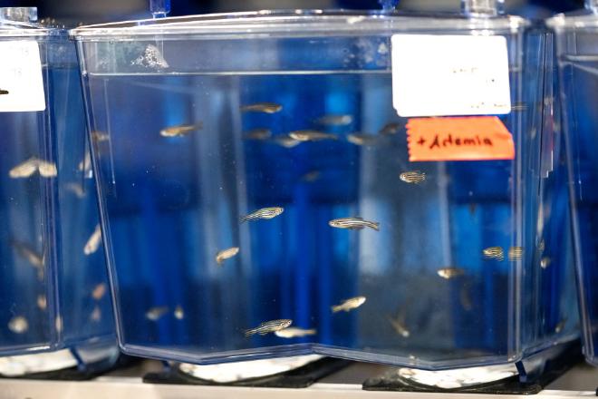
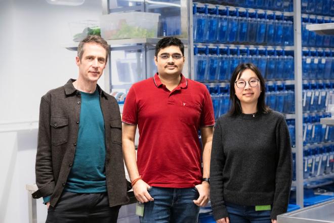
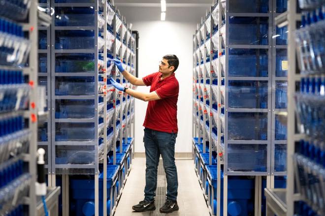
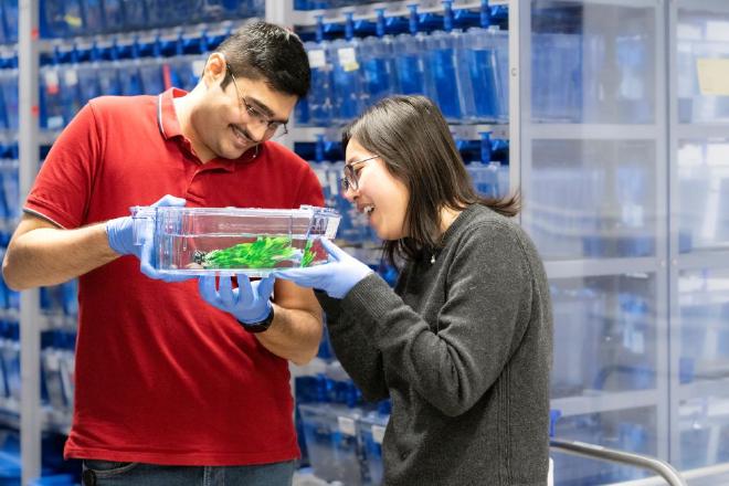
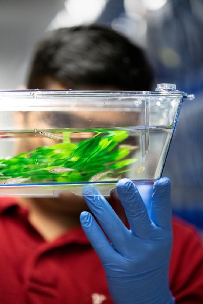
Nikhil Mishra opens a heavy door that leads into a unique room. Countless transparent boxes are stored on racks swarming with small striped fish. The water refracts through the containers, casting a bluish hue across the room. You could almost believe you were in the middle of the sea, and the gentle lapping of the water and the cozy warmth of 27 °C reinforce this feeling.
Mishra takes one of the boxes from the rack and points at a zebrafish.
“The zebrafish is an ideal organism for studying the earliest steps of development,” he explained passionately. “Their embryos are fertilized outside the mother, which means we can easily collect and study them—often hundreds at a time. They are also naturally transparent, so we can literally watch their cells divide, move, and change in real time.”
From one cell to many #
Life begins with a single fertilized egg cell, called the zygote, which begins to divide repeatedly. First into two cells, then four, then eight, and so on. This process is very similar across most species, including in humans. “Initially, these divisions happen quickly and without the cells taking on special roles. But soon, patterns begin to emerge: some cells divide more slowly, some start activating different genes, and others move to new positions,” Mishra said.
A knowledge gap #
In its early stages, the zygote depends on information provided by the mother. Only after reaching a developmental milestone called the midblastula extension (MBT) does the embryo begin to develop independently. At that point, the embryo needs to activate the appropriate genes at the right times in the correct cells. But how does it determine when and where to activate its genes? This is a fundamental question and a major knowledge gap that Mishra and the Heisenberg group at ISTA are investigating. However, they are not the only ones exploring this mystery.
ISTA’s Hannezo group is also attempting to understand how the position and timing of individual cell behavior are coordinated. These two research teams have been collaborating for some time. In particular, Yuting Irene Li, a postdoc in the Hannezo group, has greatly aided Mishra’s research with valuable expertise in theoretical physics, mathematical modeling, and statistical approaches to complex biological systems.
Geometry – the instruction manual #
This collaborative research tested a largely ignored hypothesis—that the embryo’s geometry drives its development. The ISTA scientists demonstrated that the embryo “reads” and correctly interprets the zygote’s geometry during the initial few minutes of its existence. When the researchers manipulated the early embryo geometry, it changed how cells developed later.
Think of the zygote’s geometry as an instruction manual that the embryo must read and follow as it patterns itself. If there is an error in that manual or the embryo does not read it correctly, it could lead to major problems—imagine having an intestine where your head should be.
Like a stadium wave #
Mishra explained that geometry sets off a series of highly consequential events causing cells to divide asymmetrically in an organized manner and thereby creating a gradient of cell size. These size differences create a gradient of cell cycle periods; smaller cells take longer to complete one cycle and divide into two cells.
Improving IVF outcomes #
For the ISTA scientists, the next step is to determine how universal these principles are. If similar geometric rules are also found in mammals—and especially in humans—the implications could be very significant. This is relevant as more and more people turn to assisted reproductive technologies like IVF. Even for young, healthy individuals, fewer than half of IVF embryos reach the stage where they can be implanted and lead to viable pregnancies.
In the long run, understanding these principles could help recognize early geometric “warning signs” in IVF embryos and perhaps design ways to correct or compensate for them. This could eventually contribute to more reliable embryo assessment and improved IVF outcomes.
Citations #
- The study Geometry-driven asymmetric cell divisions pattern cell cycles and zygotic genome activation in the zebrafish embryo was published in Nature Physics Authors: Nikhil Mishra, Yuting Irene Li, Edouard Hannezo & Carl-Philipp Heisenberg
Acknowledgements #
We thank N. Petridou (EMBL) for sharing results before publication. N.M. was supported by funding from the European Union’s Horizon 2020 programme under the Marie Skłodowska-Curie COFUND Actions ISTplus grant agreement number 754411. Y.I.L. acknowledges funding from the European Union’s Horizon 2020 research and innovation programme under the Marie Skłodowska-Curie grant agreement number 101034413. The research was supported by funding to C.-P.H. from the NOMIS Foundation, Project ID 1.844. We would like to thank past and present members of the Heisenberg and Hannezo groups for discussions, particularly S. Shamipour, V. Doddihal, M. Jovic, N. Hino, F. N. Arslan, R. Kobylinska and C. Camelo for feedback on the draft manuscript. This research was supported by the Scientific Service Units (SSU) of Institute of Science and Technology Austria through resources provided by the Aquatics Facility, Imaging & Optics Facility (IOF), Scientific Computing (SciComp) facility and Lab Support Facility (LSF).
Funding #
The study was supported by funding from the European Union’s Horizon 2020 programme under the Marie Skłodowska-Curie COFUND Actions ISTplus, Grant Agreement No. 754411 (NM); funding from the European Union’s Horizon 2020 research and innovation programme under the Marie Skłodowska-Curie Grant Agreement No. 101034413 (YIL) & from the NOMIS Foundation, Project ID 506 1.844 (CPH).
Information on animal studies: #
To better understand fundamental processes, for example, in the fields of neuroscience, immunology, or genetics, the use of animals in research is indispensable. No other methods, such as in silico models, can serve as an alternative. The animals are raised, kept, and treated according to strict regulations.
Contact [Notaspampeanas](mailto: notaspampeanas@gmail.com)