
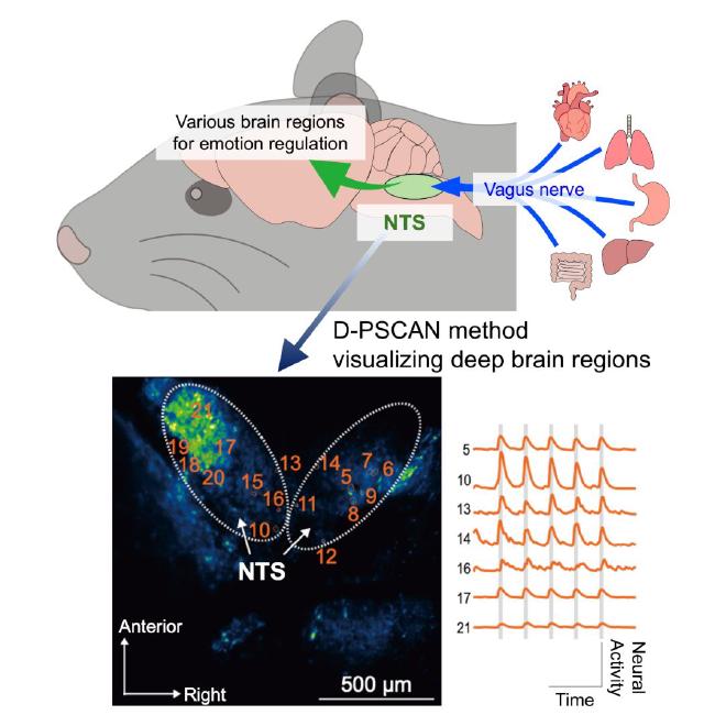
The communication between the brain and bodily organs is fundamental to emotion regulation and overall mental health. The nucleus tractus solitarii (NTS) in the brainstem is a critical hub structure mediating this interaction via the vagus nerve. Despite its importance, the NTS’s deep location has historically posed challenges for observation in living animals.
In a study published in Cell Reports Methods, the research team has developed the live NTS imaging method (named “D-PSCAN”, or Double-Prism-based brainStem imaging under Cerebellar Architecture and Neural circuits). This novel deep-brain imaging technique enables high-resolution, minimally invasive visualization of the NTS neural activity in living mice.
New Minimally Invasive Technique Unveiled. #
“One major challenge in studying the NTS is its deep location beneath the cerebellum, which has made observation in living animals difficult” explains lead author Masakazu Agetsuma. “Previously, some approaches involved removing the cerebellum to access the NTS, but this posed a major limitation: the cerebellum, major motor coordination center, has also been recognized as important for emotional regulation. Therefore, a method to observe the NTS while preserving cerebellar function has been needed.”
Detailed activity of the NTS can now be observed. #
The research team evaluated the D-PSCAN method by investigating the NTS’s response to the electrical stimulation of the vagus nerve, which conveys signals from internal organs to the NTS. They observed the specific thresholds of vagus nerve stimulation (VNS) intensity required to elicit neural responses in the NTS. They also observed that varying stimulation parameters causes distinct patterns of neural activation, including sensitization or, conversely, inhibitory effects.
Vagus nerve stimulation (VNS) has been clinically used for drug-resistant epilepsy and is currently under investigation as a treatment for depression and other psychiatric and neurological disorders. Therefore, these results highlight the potential of the D-PSCAN method for gaining valuable insights into optimizing VNS parameters for therapeutic applications.
To further investigate NTS function under more physiological conditions than electrical stimulation, the research team applied the D-PSCAN method to examine its response to the gut hormone cholecystokinin, which is naturally released after feeding. As a result, they successfully detected NTS neural activity evoked by cholecystokinin.
Future prospects #
“The brain-body interaction plays a critical role in emotion regulation, and gaining a deeper understanding of this function is expected to contribute both to the treatment of neuropsychiatric disorders and to the advancement of mental health and well-being”, says Agetsuma. “The D-PSCAN can offer a new approach to elucidate brain–body–mind interactions, and represents a valuable research tool for basic neuroscience to clinical applications.”
Funder JSPS KAKENHI, Takeda Science Foundation, Research Foundation for Opto-Science and Technology, Japan Agency for Medical Research and Development, National Institute for Physiological Sciences: Cooperative Study Program, National Center of Neurology and Psychiatry: Research Grant for Neurological and Psychiatric Disorders
- The paper Minimally invasive, wide-field two-photon imaging of the brainstem at cellular resolution was published in Cell Reports Methods. Authors: Masakazu Agetsuma ∙ Azumi Hatakeyama ∙ Daisuke Yamada ∙ Hiroshi Kuniishi ∙ Chihiro Ito ∙ Eri Takeuchi ∙ Shinji Tsuji ∙ Motosuke Tsutsumi ∙ Takako Ichiki ∙ Kohei Otomo ∙ Toshinori Yoshioka ∙ Tomoko Kobayashi ∙ Atsushi Noritake ∙ Yoshitsugu Aoki ∙ Tomomi Nemoto ∙ Hiroshi Yukawa ∙ Akiyoshi Saitoh ∙ Junichi Nabekura ∙ Masayuki Sekiguchi.