
In an article written by Emily Caldwell, from Ohio State University, she stated that discovery described in a scientific paper stems from studies in mice, in which researchers observed memory formation using advanced imaging techniques, including miniature microscopes that captured single-cell resolution in live animals.
Have you heard about dendritic compartments? #
The study shows that memories are stored in dendritic compartments: When one memory forms, the affected dendrites are primed to capture new information arriving within the next few hours, linking memories formed close in time.
“If you think of a neuron as a computer, dendrites are like tiny computers inside it, each performing its own calculations,” said lead author Megha Sehgal, assistant professor of psychology at OSU. “This discovery shows that our brains can link information arriving close in time to the same dendritic location, expanding our understanding of how memories are organized.”
The research was published in the journal Nature Neuroscience.
How we do that? #
Though most learning and memory studies have focused on how a single memory is formed in the brain, Sehgal’s lab aims to determine how we organize multiple memories.
“The idea is that we don’t form memories in isolation. You don’t form a single memory. You use that memory, make a framework of memories, and then pull from that framework when you need to make adaptive decisions,” she said.
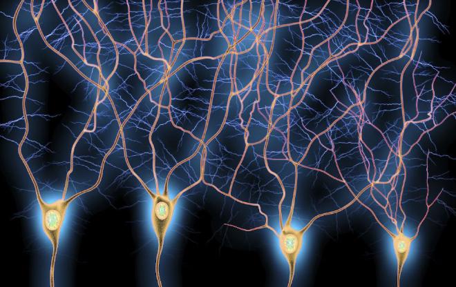
Neurons, the principal brain cells, are known to encode and relay information. Dendrites – the branch-like projections extending from neurons – serve a critical role in how information is processed, receiving incoming information and passing it to the neuronal cell body.
Dendrites stuff #
But dendrites are not just passive conduits – each dendritic branch can act as an independent computational unit. While dendrites have been thought to play an important role in the brain’s function, how they shape learning and memory has been unclear until now, Sehgal said.
The study focused on the retrosplenial cortex (RSC), a brain region crucial for spatial and contextual memory. The researchers observed that linked memories consistently engaged the same groups of RSC neurons and their dendritic branches.
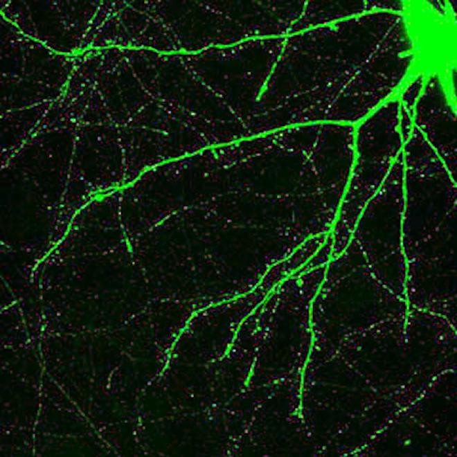
The team tracked these changes at the dendritic level by visualizing dendritic spines, tiny protrusions on dendrites where neurons communicate. The formation of new memories triggered the addition of clustered dendritic spines, a process critical for strengthening communication between neurons and facilitating learning.
Dendritic spine clusters formed after the first memory were more likely to attract new spines during a second closely timed memory, physically linking those experiences in the brain.
To confirm the role of dendrites in linking memories, the team used optogenetics, a technique that allows researchers to control neurons with light. By reactivating specific dendritic segments that had been active during memory formation, they were able to link otherwise unrelated memories, further demonstrating the importance of dendritic changes in shaping memory networks.
Memory disorders #
In addition to illuminating a previously unknown role for dendrites in linking memories, the findings open new avenues for understanding memory-related disorders, Sehgal said.
Sehgal co-led the study with Alcino J. Silva, director of the Integrative Center for Learning and Memory at UCLA, and Panayiota Poirazi, research director of the Foundation for Research and Technology-Hellas in Greece.
This work was supported by grants from the NIMH (R01 MH113071) and NIA (R01 AG013622) and from the Dr. Miriam and Sheldon G. Adelson Medical Research Foundation to A.J.S. The computational modeling work was supported by the European Commission (H2020-FETOPEN-2018-2019-2020-01, FET-Open Challenging Current Thinking, GA-863245), the National Institutes of Health (R01MH124867-01) and the Einstein Foundation Berlin to P.P. The funders had no role in study design, data collection and analysis, decision to publish or preparation of the manuscript. We acknowledge resources from the Campus Microscopy and Imaging Facility (CMIF) and the OSU Comprehensive Cancer Center (OSUCCC) Microscopy Shared Resource (MSR), The Ohio State University (RRID: SCR_025078). This facility is supported in part by grant P30 CA016058 (National Cancer Institute).
- The scientific paper Compartmentalized dendritic plasticity in the mouse retrosplenial cortex links contextual memories formed close in time was published in Nature Neuroscience. Authors: Megha Sehgal, Daniel Almeida Filho, George Kastellakis, Sungsoo Kim, Jinsu Lee, Yang Shen, Shan Huang, Ayal Lavi, Giselle Fernandes, Irene Davila Mejia, Sunaina Soans Martin, Asli Pekcan, Melody Shana Wu, Won Do Heo, Panayiota Poirazi, Joshua T. Trachtenberg & Alcino J. Silva.